The appropriate ethics declarations
The experimental protocol for the collection of bovine clinical samples was strictly in accordance with the Procedures and Guidelines for Animal Ethics in the People’s Republic of China. The animal experimental protocol was approved by the Science and Technology Ethics Committee of Ningxia University (Code: NXU-2024-003).
All methods are reported in accordance with ARRIVE guidelines.
Viruses and clinical samples
The BVDV 5’UTR gene sequence information was based on the BVDV-1 strain (MA/101/05, GenBank ID: MW054940.1) and the BoHV-1 gB gene sequence information was based on the BoHV-1 strain (BRV/2018/SMU6352, GenBank ID: MK654723.1). BVDV, BoHV-1, Bovine coronavirus (BCoV/CH/NX-GY-1/2022, GenBank ID: OQ513841.1), bovine rotavirus (BRV/CH/NX-1/2022, GenBank ID: OQ513855.1), and bovine norovirus (BNoV/CH/NX-GY-5/2022, GenBank ID: OQ430676.1), and bovine astrovirus (BAstV/CH/NX- 1/2022) used for specificity evaluation in this study were kept by the laboratory.
The clinical samples were 30 samples of cattle with respiratory disease or diarrhea syndrome collected from different farms in the Ningxia region (July 2022), including 10 nasal swabs, 10 anal swabs, and 10 serum samples. The cattle sampled were approximately 24-week-old bulls of the Simmental breed and had no history of BVDV and BoHV-1 vaccination. All samples were stored at -80 °C before being used for further testing and analysis.
Construction of standard plasmids
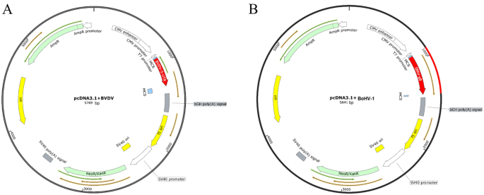
The BVDV 5’UTR gene sequence and the BoHV-1 gB gene sequence were synthesized artificially due to the limitation of the pathogen source. The whole genome sequences of BVDV and BoHV-1 were collected in GenBank, and the sequences were compared and analyzed by Mega software. The intraspecies-conserved and interspecies-specific 5’UTR (GenBank ID: MW054940.1) and gB gene (GenBank ID: MK654723.1) were selected as target genes for the RPA assay. The target genes were constructed into pcDNA-3.1 by Hind III, Xhol, and Bam HI enzymes (NEB, No. R0104/ R0146/ R0136), and transformed into E. coli recipient cells Top10, obtain pcDNA 3.1-BVDV, pcDNA 3.1-BoHV-1 recombinant plasmids.
Plasmid extraction was performed according to the Axygen Plasmid Extraction Kit (item no. AP-MN-P), and the concentration measured by Nanodrop ND-8000 spectrophotometer (Thermo Scientific, Dreieich, Germany). The DNA copy number was calculated according to the following formula: DNA copy numberâ=â(MâÃâ6.02âÃâ1023âÃâ10â9)/ (nâÃâ660), where M is the amount of DNA in nanograms, and n is length of the plasmid in bp. DNA standards were stored at â20 °C until further experiments.
Primer design
The RPA primers were manually designed based on the conserved regions of the BVDV 5’UTR gene, BoHV-1 gB gene, with reference to the instruction manual provided by the RPA reaction manufacturer Twist Dx (Cambridge, UK). Meanwhile, common PCR primers were designed for the constructed plasmid sequences (Table 2). All primers were synthesized by Sangon Biotech (Shanghai, China) and screened by observing their performance on 2% agarose gels.
Establishment of a dual RPA assay for BVDV and BoHV-1
The RPA assay was performed using the extracted recombinant plasmids pcDNA 3.1-BVDV and pcDNA 3.1-BoHV-1 as templates according to the instructions of the Twist Amp® Basic RPA kit (Twist Dx, item no. 10270-106). The following RPA reaction system was configured according to the recommendations of the kit: 2 μL of template, 11.2 μL of sterile deionized water, 29.5 μL of RPA reaction buffer, 2.4 μL each of the F and R primers at 10 pM, and 2.5 μL of magnesium acetate solution, which was mixed homogeneously and then fully solubilized in 4 mg of RPA basic lyophilisate. The RPA reaction was programmed to incubate at 39 °C for 20 min in a thermocycler (Bio Metra GmbH, 844-070-882). The amplification products were subjected to 2% agarose gel electrophoresis and the results were visualized and photographed in a UV gel imaging system.
The main parameters of the RPA assay were amplification temperature and reaction time. To optimize the BVDV, BoHV-1 dual RPA assay, experiments were performed at different reaction temperatures (30 °C, 32 °C, 35 °C, 37 °C, 39 °C, 42 °C) and reaction times (10 min, 15 min, 20 min, 25 min, 30 min, 40 min) according to the manufacturer’s recommended protocol. The optimal reaction was determined based on the specific bands in the agarose gel. To quantify the gel imaging results, the target bands (grayscale images of line regions and rectangular selections) were analyzed using ImageJ software. Intensity data were analyzed with Graphpad Prism.
Sensitivity, and specificity evaluation of BVDV and BoHV-1 dual RPA assay
The specificity of the dual RPA assays for BVDV and BoHV-1 was evaluated in bovine pathogens with similar clinical signs. BVDV, BoHV-1, BCoV, BRV, BNoV, and BAstV were detected according to established RPA assays. Nuclease-free water was used as a non-template control in the assay.
To investigate the sensitivity of the dual RPA assay for BVDV and BoHV-1, the quantified pcDNA 3.1-BVDV and pcDNA 3.1-BoHV-1 plasmids were diluted in 11 gradients, i.e., 1âÃâ1010 copies/μL to 1âÃâ100 copies/μL. The negative control template was still nucleic acid-free water.
Evaluation of the dual RPA assay using field samples
To effectively examine the effect of the dual RPA assay for BVDV and BoHV-1 established in this study on field samples, we tested 30 samples (10 nasal swabs, 10 anal swabs, and 10 sera) of bovine respiratory syndrome and diarrhea syndrome with obvious clinical symptoms in Ningxia (collected in July 2022) by RPA. The nuclease-free water was used as a negative control, the synthesized recombinant plasmids (pcDNA 3.1-BVDV and pcDNA 3.1-BoHV-1) were used as a positive control. In addition, a conventional PCR assay was performed on the same samples to compare the results with the RPA assay.
The BVDV conventional PCR primers are as follows. 5’UTR-F: TCTCGACCGGGGACATTATCT; 5’UTR-R: CATTCTGCAACGCGAAGGTG. The amplification size is 354 bp. The BoHV-1 conventional PCR primers are as follows. gB-F: GTACGACTCGTTCGCGCTCT; gB-R: CAAGTACGTCTCCAGGCTGCC. The amplification size is 481 bp.
Swab samples were generally resuspended in virus preservation solution. They are shaken for 10 min, centrifuged at 12,000 rpm for 10 min to remove solid precipitates, and filtered aseptically through a 0.22 μm membrane. Blood samples were centrifuged at 3000 rpm for 10 min after an overnight incubation at 4 °C, and the upper serum was aspirated for subsequent tests. The viral genomic DNA/RNA was extracted by the TianGen Viral Genomic DNA/RNA Extraction Kit (No. DP315) according to the manufacturer’s instructions. The RNA was reverse transcribed into cDNA by the Prime Script⢠II 1st Strand cDNA Synthesis Kit (No. 6210A) from TAKARA. The reverse transcribed cDNA and extracted DNA were subjected to RPA assay or PCR assay. The amplified products were detected by 2% agarose gel electrophoresis and quantitatively analyzed by ImageJ software.
Statistical analysis
To visually determine the specificity, sensitivity, and evaluation of dual RPA in clinical samples, we quantified the gel imaging results and analyzed the target strips (grayscale images of line regions and rectangular selections) using ImageJ software. Intensity data were analyzed with Graphpad Prism. In addition, we cropped the gel imaging results and the original full gel map imaging is shown in the Supplementary Information.


